Patient Information from ISHA – The Hip Preservation Society
Chondromatosis
Disorders Affecting the Lining (Synovium) of the Hip Joint - Chondromatosis
Hip chondromatosis and pigmented villonodular syndrome (PVNS) are both relatively rare conditions occurring in the hip joint affecting the synovial membrane (joint lining). Failure to diagnose and treat may result in further damage to the joint.
Definition
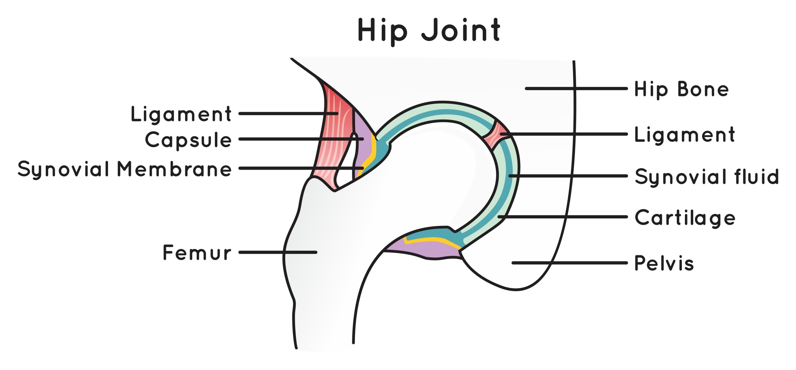
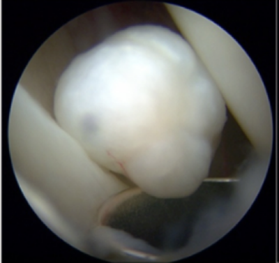
Chondromatosis is a rare, benign (non-cancerous) condition affecting the joint lining (synovium) most often in the knee but can occur in the hip joint. It generally develops between the ages of 30 and 50 and is more common in males. As the condition progresses, the lining of the affected joint grows abnormally with the development of cartilaginous nodules. These can vary in number from very few to several hundred.
There are two types of chondromatosis:
Primary chondromatosis (Reichel syndrome)
- Usually only affects one joint
- Cause is unknown
- Nodules tend to remain small resulting in less symptoms than secondary chondromatosis
Secondary chondromatosis
- Involves the formation of loose bodies resulting from joint damage as a consequence of trauma or osteoarthritis
- The size of loose bodies can vary from a few millimetres to a few centimetres
- Nodules may break off and move around the joint space further damaging the articular joint surfaces resulting in osteoarthritis
Signs and Symptoms
- Pain and tenderness
- Swelling – which can be significant
- Reduced range of movement
- Locking
- During movement there may be audible creaking, grinding or popping
Diagnosis
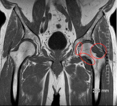
This can be difficult and may take many years. In addition to a physical examination imaging is likely to be performed but where the nodules have not calcified it may still be difficult or even impossible to see on X-ray or other imaging modalities. Examples of chondromatosis on imaging are shown adjacent.
Right: L Hip MRI image showing Chondromatosis (RNOH/Pressney, I 2024)
Surgical Treatment
This often involves removal of any loose bodies with or without removal of the joint lining – a procedure known as a synovectomy. This surgery may be performed either arthroscopically or via a open procedure using a larger incision.
Hip chondromatosis may recur in 20% of patients.
Non-Surgical Treatment
Following assessment and imaging it may be decided to monitor any symptoms or changes over time to ensure no joint damage or deterioration occurs. In some patients the condition may be self-limiting and with activity modification and use of anti-inflammatory medication and cryotherapy surgical treatment may be unnecessary. Where the condition progresses causing more severe symptoms or damage, surgical treatment may be the only option.
What to expect after surgery
Recovery following arthroscopic surgery is generally quicker than after an open procedure and hence returning to activities is also easier. Any return to sport will also depend on operative findings, and advice will be provided by the treating hip preservation surgeon and physiotherapist.
There may be limitations to weightbearing and activities during the first two or three months, which will vary amongst surgeons and will depend on operative findings and techniques performed.
Physiotherapy can begin after surgery, gradually increasing range of movement, stability, strength, mobility and function over a period of up to six months, depending on the surgery performed and individual aims.