Patient Information from ISHA – The Hip Preservation Society
Osteotomies
Osteotomies
Osteotomies are performed to avoid or delay the onset of osteoarthritis by improving joint alignment hence reducing pain and improving function. Osteotomies in hip preservation surgery involve the cutting and/or repositioning of bone around the hip joint and generally involve the pelvis, femur or tibia. The types of osteotomies described below include:
Periacetabular osteotomy (PAO)
Proximal femoral derotation
Distal tibial derotation
These are often performed when treating the following conditions which have been described in the relevant sections on this website. They include but are not limited to:
Consequences of Perthes
Avascular necrosis (AVN)
Ischiofemoral impingement
Hip dysplasia/developmental dysplasia of the hip (DDH)
Hip instability
Femoroacetabular impingement (FAI)
Rotational abnormalities of the femur and tibia

Periacetabular Osteotomy (PAO)
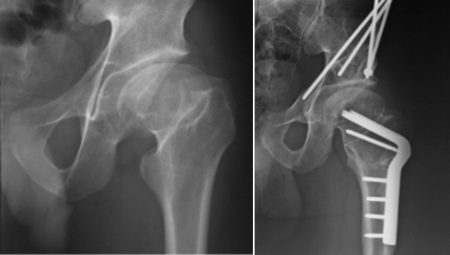
This is a type of pelvic osteotomy used to improve coverage of the femoral head by changing the orientation of the acetabulum (hip joint socket). It may also be known as a Ganz or Bernese Osteotomy. Surgery is performed under general anaesthesia and involves cutting the pelvis in a few places to free up the acetabulum (hip joint socket). The cut bones are then fixed back together with screws with the altered alignment of the socket now improving coverage of the femoral head. This helps restore stability to the hip joint and in turn improve function, reduce pain and ultimately may delay the onset of OA. Where a hip joint shows signs of osteoarthritis, a total hip replacement may become relevant.
Following a PAO there will be an extensive period of rehabilitation beginning soon after surgery and lasting several months with the ultimate aim of returning patients to normal activities including high level sports where relevant and possible. Immediately after surgery it will be necessary to use crutches for walking and weightbearing will be limited for 6-8 weeks during the initial stages of bone healing. Physiotherapy can start during this time to maintain strength and movement via exercises requiring no weightbearing. Muscles will still weaken significantly so regaining full strength and returning to full activities can take up to one year.
During the first few weeks some movements may need to be avoided and this will be explained by the treating surgeon and/or physiotherapist. If hydrotherapy facilities are accessible this may be commenced once the wounds are healed or under the direction of the treating surgeon.

Proximal Femoral Derotation
In some individuals presenting with hip and/or knee symptoms there may exist a rotational deformity (twist) in the femur. The femur may be either excessively twisted (anteversion) or less twisted than normal (retroverted), affecting hip biomechanics and may result in pain and reduced function. In order to reduce pain, improve function and avoid the early onset of joint degeneration, a proximal femoral derotation osteotomy (or varus inter-trochanteric proximal osteotomy) may be performed.
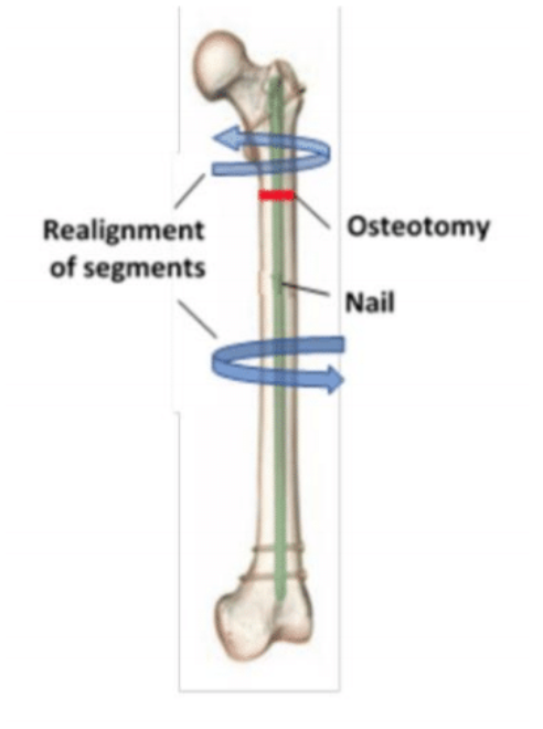
This surgical procedure, usually performed under general anaesthesia involves cutting through the upper part of the femur, rotating the top part of the femur with respect to the bottom to ensure the correct angle of the femoral neck and head, and then inserting a metal rod through the length of the femur whilst the bone heals (this rod does not necessarily need to be removed and can remain in situ).
Following a proximal femoral derotation osteotomy, there will be a lengthy period of rehabilitation with recovery and bone healing taking 6-12 months depending on the aims and objectives of the patient and the rate of bone healing. During the first few weeks weightbearing through the affected leg may be limited during which time it would be necessary to walk with crutches. This would be confirmed by the treated surgeon. It may not be necessary to wait until the bone has fully healed before resuming certain sporting activities – advice should be gained from the treating surgeon.

Distal Tibial Derotation
Hip pain may be caused by an abnormal twist (torsion) in the shin bone (tibia) causing either in-toeing (foot pointing inwards) or out-toeing (foot pointing outwards). The increased effort required to walk, run and perform other activities whilst trying to keep foot pointing forwards can lead to either knee and/or hip pain. This torsion may cause incorrect alignment of the hip joint and a surgical procedure involving an osteotomy may be performed to correct this. This procedure aims to restore normal alignment and hence improve function and reduce pain.
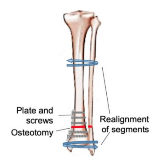
Surgery which takes place under general anaesthesia involves an osteotomy (cutting) through the lower part of the tibia and fibula, just above the ankle. Alignment is corrected by rotating the shin bone to improve alignment. The bones are then stabilised using a titanium plate and screws. This metalwork may not need to be removed and can remain in situ. A plaster cast is usually applied for around two weeks during which time no weight can be applied through the leg. Mobilising with crutches will be necessary. Once the wound and bone healing has been assessed after two weeks it may be possible to gradually return to walking normally under the guidance of the treating surgeon and physiotherapist.